
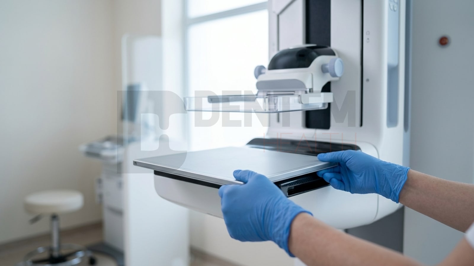
A large number of analog mammography units are still in daily service across radiology departments. The generator is healthy, the tube has life left in it, the C-arm and compression system are mechanically sound. The problem is not the machine itself: it is film supply, chemical processing, physical archiving and the reporting chain around it. A new digital mammography system, meanwhile, is a six-figure investment.
Mammography retrofit is the middle path. It replaces the image receptor, the film cassette, with a digital flat panel detector. Everything else stays where it is.
There are two ways to do this, and the difference between them decides the duration and cost of the project. Below we cover that distinction first, then the technical detail.
Two Routes: Cassette Size or Integrated Detector
1. Cassette-size detector. The detector housing matches the standard dimensions of a film cassette. You take the cassette out and slide the detector into the same slot. The gantry is not dismantled, the bucky assembly is untouched, and no permanent mechanical change is made to the unit. The Mammo 1012C is in this class: its housing conforms to ISO 4090, and it works with the unit’s existing AEC system.
2. Integrated detector. The bucky assembly is removed together with the ion chamber, a detector tray is built in its place, and the AEC is re-interfaced. More work, longer installation, but a higher technical ceiling. The Mammo 1012F and Mammo 1012P fall into this class.
In practice most projects are solved by the first route. Because the result is achieved without touching the machine, downtime is shorter and cost is lower, the cassette-size detector is the default choice. The second route comes up when the facility wants to move to tomosynthesis or needs higher resolution.
What Is a Mammography Retrofit?
A retrofit keeps the side of the unit that generates X-rays and positions the breast, and converts the side that captures the image. The result is a system that produces full field digital mammography (FFDM).
| Stays in place | Gets replaced |
|---|---|
| X-ray tube and generator | Film cassette |
| C-arm, gantry, compression paddle | Image receptor (flat panel detector) |
| Cassette slot and bucky (cassette-size route) | Processing, darkroom, film archive |
| Existing AEC (cassette-size route) | Image processing software and PACS link |
| Lead-shielded room and installation | Reporting and archiving workflow |
The image appears on screen within seconds of exposure. Film, chemistry and physical archiving are removed from the workflow entirely.
When a Retrofit Makes Sense, and When It Does Not
To be straightforward about it, a retrofit is not the right decision for every unit.
It makes sense when:
- The generator and tube are healthy, with at least five years of expected life
- The mechanics (compression, rotation, height) work without fault
- Annual exam volume does not justify a new system purchase
- The facility wants out of film supply and archiving
- Building, services or room dimensions cannot accommodate a new gantry
It does not make sense when:
- Tube output has dropped, or the generator fails repeatedly
- The manufacturer has ended support and spare parts are unobtainable
- The facility plans to move to digital breast tomosynthesis (DBT) soon and the existing gantry cannot perform the tomosynthesis sweep
- Compression force calibration will not hold
This assessment cannot be made remotely. It is made at the machine, by measuring generator output and observing AEC behaviour.
AEC: The Critical Point
If one technical subject decides whether a retrofit succeeds, it is automatic exposure control.
In analog mammography, the AEC ion chamber sits behind the cassette. The beam passes through the breast and the film, reaches the chamber, and once the chamber has collected enough signal it tells the generator to terminate the exposure.
In the cassette-size route that assembly stays in place. The Mammo 1012C is designed to work with the unit’s own AEC system, and it also carries an AED (Automatic Exposure Detection) function that senses the X-ray beam through a dedicated sensor and triggers the detector itself. The generator’s electrical circuitry is not modified.
In the integrated route the bucky is removed together with the ion chamber. The detector that takes its place must both produce the image and provide the exposure termination signal. This is solved either by detector-based AED, or by an interface board that converts the detector signal into the format the generator’s AEC input expects.
What happens if it is done badly? The exposure terminates early and the image comes out noisy, or it terminates late and the patient receives unnecessary dose. Either way the view has to be repeated. A repeat exposure is the most expensive error in mammography: it costs dose, patient comfort and screening programme throughput at the same time.
That is why the first question to ask about a retrofit quote is not “how many pixels does the detector have” but “how will the AEC work, and who will calibrate it”.
Chest Wall Distance: The Tissue You Lose
Mammography has to image the tissue closest to the chest wall. The gap between the edge of the detector’s active area and the physical edge of its housing is tissue that simply cannot be imaged.
The Mammo 1012C and Mammo 1012F both hold this distance at 3 mm. The Mammo 1012P uses a tapered housing profile with a super slim edge to extend the field of view towards the chest wall.
When selecting a retrofit detector, this dimension matters as much as pixel pitch. It is also one of the main reasons a general purpose radiography detector cannot be used for mammography.
Pixel Pitch and Microcalcifications
What separates mammography from other radiography applications is the size of the finding being looked for. Microcalcifications are often under half a millimetre, and they are the most valuable sign of early stage breast cancer. Seeing them demands very high spatial resolution.
Pixel pitch is directly tied to the theoretical resolution limit (Nyquist):
- 85 µm pixel pitch gives roughly 6 lp/mm. This is sufficient for standard FFDM screening and diagnosis. The Mammo 1012C and 1012F sit here.
- 49.5 µm pixel pitch gives roughly 10 lp/mm. This is a clear advantage for fine microcalcification clusters and for tomosynthesis. The Mammo 1012P is in this class.
Smaller pixels are not automatically better: as the pixel shrinks, the same signal falls over less area and noise rises. Sensor technology and scintillator efficiency therefore become critical in high resolution detectors.
The iRay Mammo Series Compared
| Specification | Mammo 1012C | Mammo 1012F | Mammo 1012P |
|---|---|---|---|
| Retrofit method | Slides into cassette slot | Integrated mounting | Integrated mounting |
| Cassette standard | ISO 4090 | None | None |
| Sensor | Amorphous Silicon | Amorphous Silicon | CMOS |
| Pixel pitch | 85 µm | 85 µm | 49.5 µm |
| Resolution | 6 lp/mm | 6 lp/mm | 10 lp/mm |
| Active area | 235 × 290 mm | 239 × 300 mm | 10″ × 12″ |
| Chest wall distance | ≤ 3 mm | 3 mm | Slim edge design |
| Data interface | GigE | Wired | 10G Ethernet |
| Trigger | Software / AED | AED | Software / AED |
| Works with existing AEC | Yes | Interface required | Interface required |
| Application | FFDM | FFDM | FFDM + DBT |
| Weight | ≈1.3 kg | 4 kg | 2 kg |
How to choose? If the goal is to convert the existing analog unit to digital in the shortest time and with the least intervention, the 1012C is the right answer, and most projects are solved this way. The 1012P comes into play when the facility is considering tomosynthesis later, or when resolving fine calcifications in dense breast tissue is a priority.
Compatibility is not determined by brand name but by cassette slot dimensions, generator communication interface and AEC type. Most of the analog mammography bodies encountered in the field are suitable for retrofit, but every unit is assessed on its own site survey.
Dose: What to Expect After a Retrofit
Dose reduction is the most talked about benefit of going digital, but it does not happen automatically. Dose depends on AEC calibration and exposure protocol as much as on detector quantum efficiency.
EUREF criteria provide the reference: average glandular dose (AGD) on a 45 mm PMMA phantom should be under 2.5 mGy. That is the value to measure at acceptance testing after a retrofit. Without that measurement, claiming “our dose went down” has nothing behind it.
Other factors that affect dose:
- AED / AEC calibration: must be verified separately across different breast thicknesses and densities
- Correct collimation: the beam field should be limited to the detector active area
- Periodic quality control: only regular calibration shows that dose output has not drifted over time
For the full picture on the detector side, see our guide on wired versus wireless flat panel detectors.
Image Processing, DICOM and PACS
A retrofit is not only a hardware change. Without the right software chain behind the detector, what you have instead of film is simply a raw image file.
The chain covers:
- Mammography-specific image processing: breast tissue has a wide dynamic range, and general radiography processing algorithms fall short here
- DICOM MG modality: images must be produced under the mammography modality with correct laterality (right/left) and view (CC/MLO) tags
- Worklist integration: patient data arrives automatically from the HIS, removing manual entry errors
- PACS archiving: access to prior studies for comparison reading is a basic requirement of screening programmes
The Mammo series exports images in DICOM format to PACS, CD/DVD or external storage.
Regulation and Acceptance Testing
Changing the image receptor is a recorded change to the device. The administrative side of a retrofit covers:
- Updating the radiation licence record so that the licensed device entry reflects the new configuration
- Device registry: the installed detector must be registered and its unique device identifier passed to the facility
- Acceptance testing: AGD, contrast to noise ratio (CNR), spatial resolution and uniformity are measured after commissioning
- Periodic quality control: the acceptance test values become the reference against which later measurements are compared
Required documentation and test scope vary by facility type and jurisdiction, so we recommend confirming the applicable requirements before the project starts.
The Retrofit Process on Site
- Site survey: generator output, tube condition, cassette slot dimensions and AEC type are established on site
- Feasibility report and quote: which detector fits, which route is needed, and total cost
- Installation: in the cassette-size route the detector goes into the slot and the workstation is set up. In the integrated route the bucky and ion chamber are removed, and the detector tray and interface are fitted
- AEC calibration: the exposure curve is established across a range of phantom thicknesses
- Acceptance testing and QC: measurements are taken and reference values recorded
- Training and go-live: technologist and radiologist training is delivered, the PACS workflow goes live
A retrofit done with a cassette-size detector usually takes the unit out of service for less than a day. The integrated route can extend that to a few days.
Frequently Asked Questions
Can my analog mammography unit be converted to digital?
If the generator and mechanics are healthy, most likely yes. A definite answer comes after a site survey. What decides it is not the brand but cassette slot dimensions, generator interface and AEC type.
Does the machine have to be dismantled?
Not if a cassette-size detector is used. The Mammo 1012C conforms to the ISO 4090 cassette standard, so it goes straight into the existing cassette slot and works with the unit’s own AEC. Dismantling only applies to detectors that require integrated mounting.
Retrofit or a new digital mammography system?
A retrofit sits well below the cost of a new FFDM system and uses your existing room as it is. However, if your generator is at the end of its life, the retrofit investment will come back around before long. The decision follows the remaining life of the machine.
I have a CR system. Do I still need a retrofit?
CR (cassette reader) systems are digital, but the workflow still depends on carrying cassettes and generally requires higher dose. A DR retrofit removes the cassette cycle completely and brings the image to screen within seconds.
Can I do tomosynthesis (DBT)?
On the detector side, the Mammo 1012P supports tomosynthesis. However, DBT also requires that the gantry can perform the tube sweep. A classic analog gantry cannot, so tomosynthesis is only possible on suitable bodies.
Will dose really drop after a retrofit?
It is expected to, but it is not guaranteed. AEC calibration is what decides it. At acceptance testing, average glandular dose measured on a 45 mm PMMA phantom should be under the EUREF limit of 2.5 mGy.
Conclusion
A mammography retrofit is the way to move to a digital workflow without scrapping a working machine. Done properly, the facility is freed from film and archiving, reporting time drops, images flow into PACS and dose comes under control.
With a cassette-size detector, that transition is completed without touching the body of the machine at all. The difference is not the numbers on the detector label. It is whether the team doing the integration understands what is specific to mammography.
As iRay’s distributor in Türkiye, Dentem Health handles every step of a mammography retrofit in one place: site survey, detector supply, AEC integration, acceptance testing and training.
Free feasibility survey for your unit: WhatsApp: +90 533 604 36 60 · Phone: +90 (216) 504 00 47