
Mammography Digital Conversion: Go Digital Without Replacing Your Analog Unit
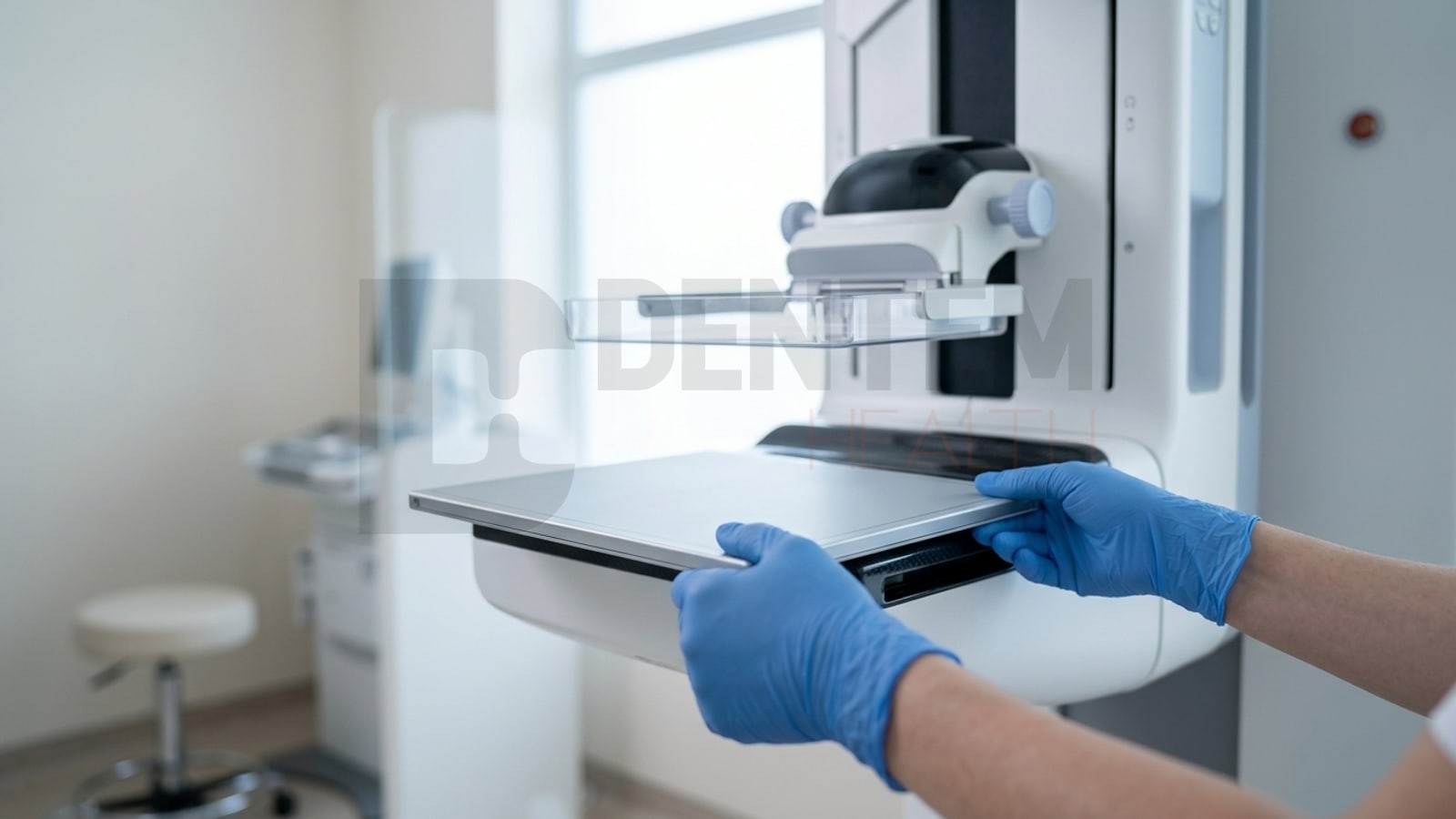
The generator, C-arm and compression system stay in place. A digital flat panel detector takes the place of the film cassette.
Can an analog mammography unit be converted to digital?
A unit whose cassette slot and automatic exposure control (AEC) are suitable can go digital with a cassette-size digital detector, without modifying the unit. The site survey decides suitability. With the iRay Mammo 1012 series, Dentem carries out the site survey, installation, AEC integration, acceptance test and training with the same team.
The decision is made not by brand name but by cassette slot size, generator interface and AEC type.
Which units does it apply to?
Three things are checked at the survey: cassette slot size (ISO 4090), AEC type, and generator and triggering. There are two methods.
Cassette-size detector
The detector goes into the same slot as the film cassette. The gantry is not dismantled, the bucky assembly is left alone and the unit’s own AEC system is used. The Mammo 1012C is in this class.
Integrated detector
The bucky assembly is removed together with the ion chamber, a detector tray is built and the AEC is interfaced again. The Mammo 1012F and Mammo 1012P are in this class.
When does a retrofit make no sense?
- If the tube output has dropped or the generator breaks down repeatedly
- If the manufacturer has ended support and spare parts cannot be found
- If the institution plans to do digital breast tomosynthesis (DBT) soon and the gantry does not support the tomosynthesis movement
- If the compression force calibration does not hold
This assessment cannot be made remotely; it is made at the unit.
Process
-
1
Site survey
Generator output, tube condition, cassette slot size and AEC type are established at the unit.
-
2
Compatibility check
Which detector fits and which method is needed is settled in writing.
-
3
Supply
Dentem supplies the chosen iRay Mammo detector and the workstation.
-
4
Installation
With a cassette-size solution, the detector goes into the existing slot. With an integrated solution, the bucky and ion chamber are removed and a detector tray is built.
-
5
AEC integration
The exposure curve is set for different phantom thicknesses.
-
6
Acceptance test
Measurements are taken and reference values are recorded.
-
7
Training
Technologists and radiologists are trained and the PACS workflow goes live.
Acceptance test scope
After commissioning, the average glandular dose (AGD), contrast-to-noise ratio (CNR), spatial resolution and uniformity are measured. The acceptance test scope is given in writing after the site survey.
Permit and license applications are made by the institution that operates the device; Dentem prepares the device information and documents and guides you.
Dose and image quality
The EUREF criteria are used as the reference: in a 45 mm PMMA phantom the average glandular dose (AGD) must be below 2.5 mGy. This is the value to be measured at the acceptance test after a retrofit.
Dose depends on AEC calibration and the exposure protocol as much as on the detector; a reduction is not guaranteed, it is measured.
PACS, DICOM and HIS
Images from Mammo detectors can be transferred in DICOM format to PACS, to CD/DVD, to USB memory or to an external disk. The chain that is set up covers mammography-specific image processing, correct laterality (right/left) and view (CC/MLO) labels, patient information from the hospital information system (HIS) by worklist, and PACS archiving for comparative reading.
Technical data
For the iRay Mammo 1012 series, the values are the same as the specifications on the product pages.
| Feature | Mammo 1012C | Mammo 1012F | Mammo 1012P |
|---|---|---|---|
| Detector type | Amorphous Silicon | Amorphous Silicon | CMOS |
| Pixel pitch | 85 µm | 85 µm | 49.5 µm |
| Resolution | 6 lp/mm | 6 lp/mm | 10 lp/mm |
| Active area | 235 × 290 mm (10″ × 12″) | 10″ × 12″ (239 × 300 mm) | 10″ × 12″ |
| Connection | GigE | Wired | 10G Ethernet |
| Weight | ~1.3 kg | 4 kg | 2 kg |
In the same survey: moving from CR to DR
If you have a working generator, only the imaging chain changes: the flat panel detector, the workstation software and a PACS or printer connection. The brand and model of your X-ray unit are enough; at the survey we check compatibility, install and train.
Flat panel detectorsFrequently Asked Questions
Can an analog mammography unit be converted to digital?
A unit whose cassette slot and automatic exposure control (AEC) are suitable can go digital with a cassette-size digital detector, without modifying the unit. The site survey decides suitability. With the iRay Mammo 1012 series, Dentem carries out the site survey, installation, AEC integration, acceptance test and training with the same team.
Does the unit have to be dismantled?
Not if a cassette-size detector is used. Because the Mammo 1012C conforms to the ISO 4090 cassette standard, it goes straight into the existing cassette slot and works with the unit’s own AEC system. Dismantling only applies to detectors that need an integrated mount.
Does the dose really go down after a retrofit?
It is expected to, but this is not guaranteed. AEC calibration is the deciding factor. At the acceptance test, the average glandular dose measured in a 45 mm PMMA phantom must be below the EUREF limit of 2.5 mGy.
Can I do tomosynthesis (DBT)?
On the detector side, the Mammo 1012P supports tomosynthesis. For DBT, however, the gantry must also be able to make the tube swing movement. A classic analog gantry cannot make this movement, so tomosynthesis is only possible on suitable units.
Request a site survey
Send us the brand and model of your unit and we will check suitability together at the site survey.