
Progress on the hardware side of flat panel detectors is slowing down. Pixel pitch, scintillator efficiency and A/D conversion have reached a certain maturity. The real gains are no longer produced in the panel itself, but in the layer that processes the raw data the panel delivers.
The four technologies iRay has introduced recently work in exactly that layer: iGrid, iNS, iPositioning and iTomo. All four are software and algorithm solutions that sit on top of existing detector hardware. Below we cover what each one does, which problem it answers, and what it changes in practice.
1. iGrid: Scatter Suppression Without a Physical Grid
In radiography, scattered radiation is the single largest factor degrading image contrast. The classic answer is to place an anti-scatter grid between the patient and the detector. A physical grid carries three real costs, however: it absorbs part of the usable dose (so more dose has to be delivered to the patient), it adds cost, and it can leave grid line artefacts in the image.
iGrid is a physics-based technique that suppresses scatter by calculating it rather than blocking it with a grid. It estimates the scatter distribution from the raw image and subtracts it.
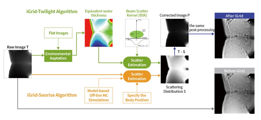 The two iGrid algorithms. Image: iRay Technology
Two different algorithms are used:
- iGrid-Twilight: performs environmental adaptation from flat images, derives the equivalent water thickness, computes the Beam Scatter Kernel (BSK) and arrives at a scatter estimate.
- iGrid-Sunrise: produces a scatter estimate from model-based, offline Monte Carlo simulations together with a specified body position.
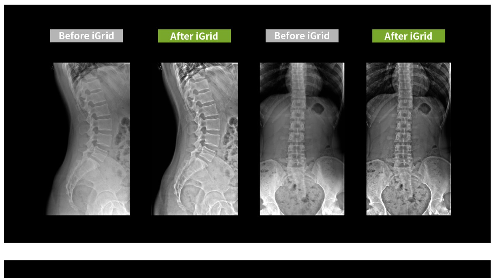
Both paths produce a scatter distribution S. Subtracting S from the raw image T gives the corrected image. The post-processing applied afterwards is identical on both images, which makes the comparison a fair one.
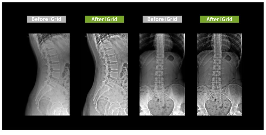 Before and after iGrid. Image: iRay Technology
What does it deliver? The four headings iRay puts forward: lower dose, lower cost, more convenient application and higher image quality. The grid line artefact problem also disappears.
Who benefits? Any scenario where carrying a physical grid is awkward. Mobile radiography and screening vehicles in particular, because there a grid means both weight and alignment trouble. Alongside human medicine, iRay also lists veterinary, industrial and mobile DR applications. For the detector side of mobile work, see our portable X-ray selection guide.
2. iNS: Noise Suppression With a Neural Network
When dose is lowered, the first thing to degrade is not contrast but quantum noise. Fewer photons means a noisier image. Classic noise filters soften the problem, but they do it by blurring: fine detail leaves along with the noise.
iNS (Intelligent Noise Suppression) aims to break that trade-off. A convolutional neural network learns the physical noise model of the detector and removes quantum noise without losing detail.
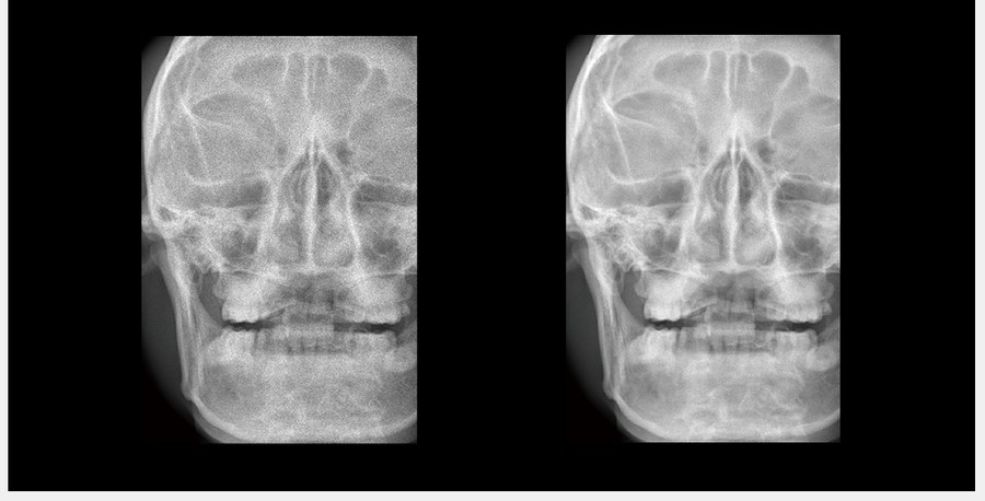 Before and after iNS. Image: iRay Technology
The measurable side of the claim:
| Measurement | Before iNS | After iNS |
|---|---|---|
| Mean | 3472.103 | 3470.940 |
| Standard deviation | 34.243 | 23.167 |
| SNR | 101.40 | 149.82 |
The point to notice is that the mean barely moves. The signal stays where it was; only the standard deviation, that is the noise, comes down. Signal flat, noise down, signal to noise ratio up.
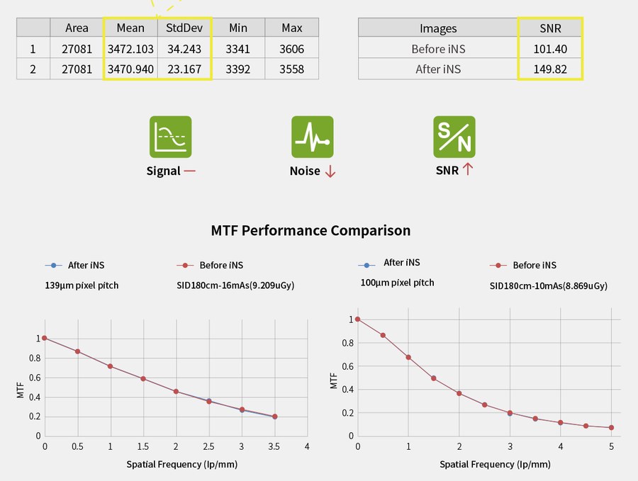 SNR values and MTF comparison. Image: iRay Technology
The genuinely critical chart is the MTF comparison underneath. Across two different test conditions (139 µm pixel pitch, SID 180 cm, 16 mAs / 9.209 µGy and 100 µm pixel pitch, SID 180 cm, 10 mAs / 8.869 µGy) the MTF curves before and after iNS lie on top of each other. Spatial resolution is preserved.
That is the evidence behind the “non-destructive denoising” claim: a filter that blurred would push the MTF curve down.
Who benefits? Anyone running low dose protocols. Paediatrics, screening programmes and repeated follow-up exposures are where dose is discussed most.
3. iPositioning: AI-Assisted Positioning
The most common cause of a repeat exposure in radiography is not equipment failure but incorrect positioning and the wrong dose setting. iPositioning is an AI-based human-machine interaction application that aims to hand both of those errors to the machine.
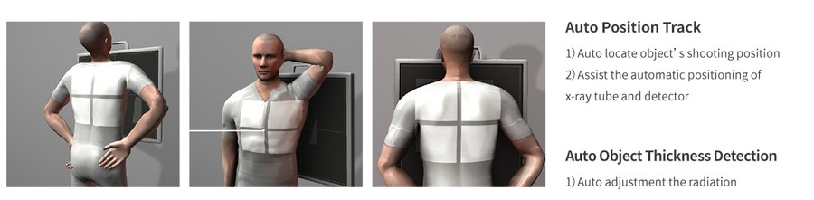 Automatic position tracking and object thickness detection. Image: iRay Technology
It has two functions:
- Auto Position Track: tracks the patient’s shooting position and assists in the automatic positioning of the detector and the X-ray tube. iRay positions this directly as a saving in labour costs.
- Auto Object Thickness Detection: uses a beam limiter with an integrated TOF (Time of Flight) module. It automatically identifies the body thickness of the patient or animal and adjusts the exposure dose accordingly.
Who benefits? High volume departments and sites running with a single technologist. Veterinary radiology too, where holding the patient in the right position is a problem in its own right.
4. iTomo: Limited-Angle Tomography at Low Dose
The fundamental limit of two-dimensional radiography is tissue superposition: overlapping structures can hide a lesion, or create the impression of one that is not there. Computed tomography solves this, but at several times the dose and the cost.
iTomo is a method for performing high-resolution limited-angle tomography at radiation dose levels comparable to projection radiography. A projection series is acquired while the tube travels along an arc, and is then reconstructed into slices.
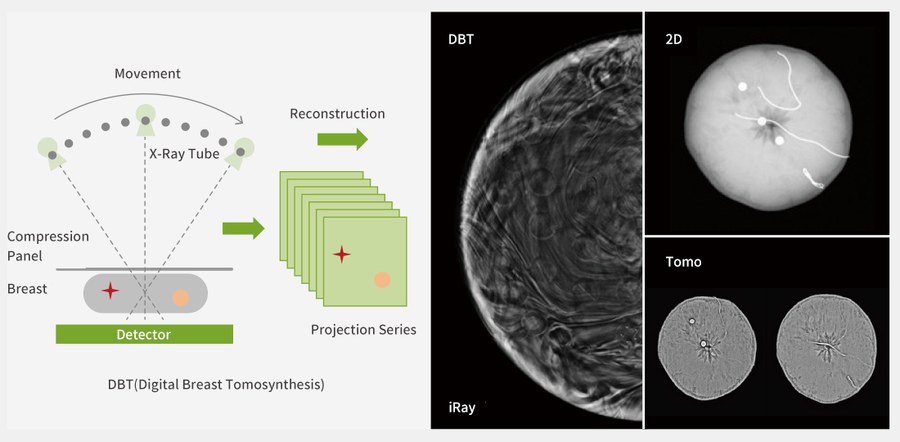 The DBT principle, projection series and reconstruction. Image: iRay Technology
Its best known application is digital breast tomosynthesis (DBT). With the breast under compression, the tube sweeps through an arc, a projection series is collected and slices are produced. Lesions that 2D mammography misses in dense breast tissue can separate out in the slices.
Tomosynthesis matters separately on the mammography side: if you are considering converting an existing analog unit to digital, our mammography retrofit guide explains when tomosynthesis is possible and when it is not.
What the Four Have in Common
All four look in the same direction: more diagnostic information, from less hardware and less dose.
- iGrid replaces a physical part (the grid) with software
- iNS pays the price of low dose (noise) in software
- iPositioning cuts repeat exposures by reducing human error
- iTomo partly solves the blindness of 2D without going to CT dose
This is also an indicator of where the detector market is heading. Judging a panel on pixel pitch and DQE alone is now incomplete; what the processing chain behind it does is at least as decisive.
Which Detector Has Which?
These technologies are presented at platform level and may not be available in the same form on every detector model. Whether a given model supports one of them has to be confirmed against that unit’s current software version.
As iRay’s distributor in Türkiye, Dentem Health can assess with you which of these your existing installation could support.
Technology assessment for your equipment: WhatsApp: +90 533 604 36 60 · Phone: +90 (216) 504 00 47